Med-Fit Tech Assistant
Human Anatomy - Learning Module #3
The Neuro-Muscular System
Part B
Part B: Muscle Histology & Physiology
Basic Structure and Function of Skeletal Muscle
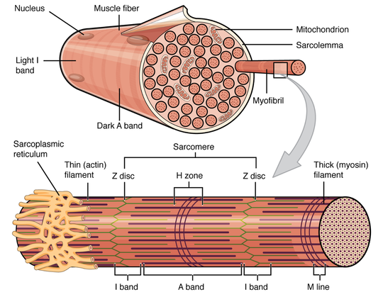
A skeletal muscle is a highly organized tissue that follows a graduated bundling system:
Basic Structure and Function of Skeletal Muscle
A skeletal muscle is a highly organized tissue that follows a graduated bundling system:
- Each muscle is composed of many fascicles.
- Each fascicle is composed of many muscle fibers.
- Each muscle fiber (the cellular unit) is composed of many myofibrils.
- Each myofibril is composed of many actin (thin) and myosin (thick) filaments.
The graduated bundles are each surrounded by their own connective tissue:
- Each muscle is surrounded by its epimysium.
- Each fascicle is surrounded by its perimysium.
- Each muscle fiber (cell) is surrounded by its endomysium.
- The cellular membrane of a muscle fiber (cell) is called its sarcolemma.
- Each myofibril within the muscle fiber (cell) is surrounded by the intra-cellular sarcoplasmic reticulum (SR).
The thin (actin) and thick (myosin) filaments within the muscle fiber (cell) are strictly arranged in an overlapping pattern that gives skeletal muscle tissue a striated appearance under a microscope. The repeating pattern unit is called a sarcomere.
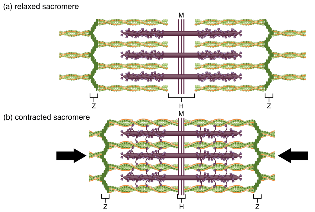
The contraction and relaxation (elongation) of a muscle is accomplished by collectively and simultaneously sliding the actin (thin) and myosin (thick) filaments in all the fibrils in all the muscle fibers in the fascicles of the muscle that get activated by their motor neurons.
The contraction and relaxation (elongation) of a muscle is accomplished by collectively and simultaneously sliding the actin (thin) and myosin (thick) filaments in all the fibrils in all the muscle fibers in the fascicles of the muscle that get activated by their motor neurons.
Both neurons and skeletal muscle cells are electrically excitable, meaning that they are able to generate action potentials. An action potential is a special type of electrical signal that can travel along a cell membrane as a wave. This allows a signal to be transmitted quickly over long distances.
Cross-Bridge Cycling
During contraction the myosin heads of the thick filament bind to actin and pull the thin filament which shortens the sarcomere and produces force. However, the length of the myosin hinge region allows each myosin head to only pull a very short distance before it must reset to pull again. For thin filaments to continue to slide past thick filaments during muscle contraction, myosin heads must pull the actin at the binding sites, detach, re-cock, attach to more binding sites, pull, detach, re-cock, etc. This repeated molecular movement to cause muscle contraction is called cross-bridge cycling and is dependent on ATP (Adenosine Tri-Phosphate).
Sources of ATP
ATP supplies the energy for muscle contraction to take place. It also provides the energy for the active-transport Calcium pumps in the sarcoplasmic reticulum necessary for contraction.
Muscle contraction does not occur without sufficient amounts of ATP. ATP is a relatively unstable molecule and storing large amounts for any amount of time is not possible. Because the amount of ATP stored in muscle is very low and is sufficient to power only a few seconds worth of contractions. ATP must be regenerated and replaced quickly to allow for sustained contraction.
There are three mechanisms by which ATP can be regenerated:
Aerobic respiration is much more efficient than anaerobic glycolysis, but it cannot be sustained without a steady supply of oxygen to the skeletal muscle. It is also much slower. To compensate, muscles store a small amount of excess oxygen in proteins called myoglobin, allowing for more efficient muscle contractions and less fatigue. Aerobic training also increases the efficiency of the circulatory system so that oxygen can be supplied to the muscles for longer periods of time.
Muscle Tension
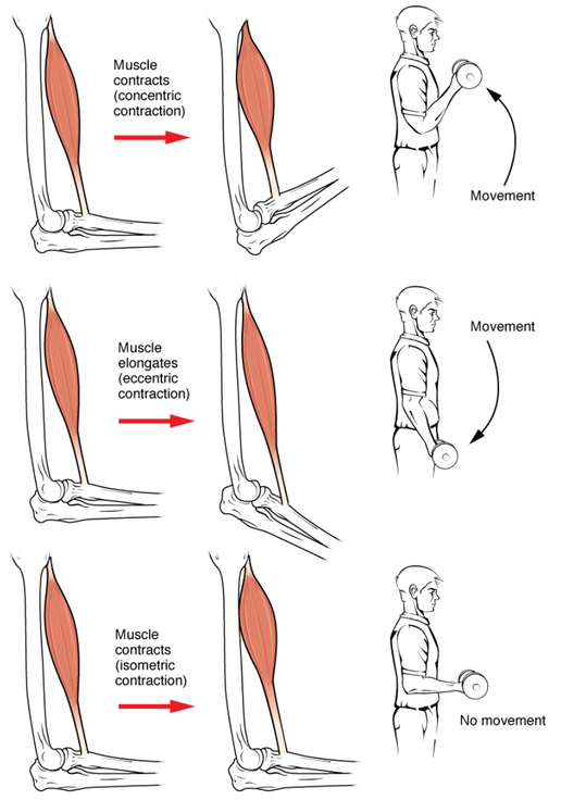
To move an object, referred to as a load, the muscle fibers of a skeletal muscle must shorten. The force generated by a contracting muscle is called muscle tension. Muscle tension can also be generated when the muscle is contracting against a load that does not move, resulting in two main types of skeletal muscle contractions: isotonic contractions (both concentric and eccentric) and isometric contractions.
Cross-Bridge Cycling
During contraction the myosin heads of the thick filament bind to actin and pull the thin filament which shortens the sarcomere and produces force. However, the length of the myosin hinge region allows each myosin head to only pull a very short distance before it must reset to pull again. For thin filaments to continue to slide past thick filaments during muscle contraction, myosin heads must pull the actin at the binding sites, detach, re-cock, attach to more binding sites, pull, detach, re-cock, etc. This repeated molecular movement to cause muscle contraction is called cross-bridge cycling and is dependent on ATP (Adenosine Tri-Phosphate).
Sources of ATP
ATP supplies the energy for muscle contraction to take place. It also provides the energy for the active-transport Calcium pumps in the sarcoplasmic reticulum necessary for contraction.
Muscle contraction does not occur without sufficient amounts of ATP. ATP is a relatively unstable molecule and storing large amounts for any amount of time is not possible. Because the amount of ATP stored in muscle is very low and is sufficient to power only a few seconds worth of contractions. ATP must be regenerated and replaced quickly to allow for sustained contraction.
There are three mechanisms by which ATP can be regenerated:
- Creatine Phosphate metabolism can power the first 15 seconds of muscle contraction.
- Anaerobic Glycolysis is a non-oxygen-dependent process to breakdown glucose (sugar) to produce ATP. The sugar used in glycolysis can be provided by blood glucose or by metabolizing glycogen that is stored in the muscle. This metabolic pathway can power muscle contraction for about 1 minute. More stores of glycogen are available in the liver, which can be metabolized and released as glucose into the blood.
- Aerobic Respiration is an oxygen dependent process that breaks down glucose or other nutrients to produce carbon dioxide, water, and ATP. Approximately 95 percent of the ATP required for resting or moderately active muscles is provided by aerobic respiration, which takes place in mitochondria.
Aerobic respiration is much more efficient than anaerobic glycolysis, but it cannot be sustained without a steady supply of oxygen to the skeletal muscle. It is also much slower. To compensate, muscles store a small amount of excess oxygen in proteins called myoglobin, allowing for more efficient muscle contractions and less fatigue. Aerobic training also increases the efficiency of the circulatory system so that oxygen can be supplied to the muscles for longer periods of time.
Muscle Tension
To move an object, referred to as a load, the muscle fibers of a skeletal muscle must shorten. The force generated by a contracting muscle is called muscle tension. Muscle tension can also be generated when the muscle is contracting against a load that does not move, resulting in two main types of skeletal muscle contractions: isotonic contractions (both concentric and eccentric) and isometric contractions.
In isotonic contractions, where the tension in the muscle stays relatively constant, a load is moved as the length of the muscle changes.
An isometric contraction occurs when a muscle produces tension, but there is no change in the length of the muscle. Isometric contractions involve sarcomere shortening and increasing muscle tension, but do not move a load, as the force produced cannot overcome the resistance provided by the load or stationary object. In everyday living, isometric contractions are active in maintaining posture and maintaining bone and joint stability.
Most actions of the body are the result of a combination of isotonic and isometric contractions working together to produce a wide range of outcomes. These muscle activities are under the control of the nervous system. A crucial aspect of nervous system control of skeletal muscles is the role of motor units (see part A).
Muscle Strength
The number of skeletal muscle fibers in a given muscle is genetically determined and does not change. Muscle strength is determined by the amount of myofibrils and sarcomeres within each fiber. Factors, such as exercise and hormones, can increase the production of sarcomeres and myofibrils within the muscle fibers to cause a change in the muscle called hypertrophy, which is an increase in mass (size, bulk) of skeletal muscle (but not the number of muscle fibers).
Likewise, decreased use of a skeletal muscle results in atrophy, where the number of sarcomeres and myofibrils decrease (but not the number of muscle fibers). It is common for a limb in a cast to show atrophied muscles when the cast is removed after just a few weeks, and certain diseases, such as polio, cause muscles to atrophy.
- Concentric contraction involves tension that shortens the muscle to move a load.
- Eccentric contraction involves tension while the muscle lengthens as the load moves.
An isometric contraction occurs when a muscle produces tension, but there is no change in the length of the muscle. Isometric contractions involve sarcomere shortening and increasing muscle tension, but do not move a load, as the force produced cannot overcome the resistance provided by the load or stationary object. In everyday living, isometric contractions are active in maintaining posture and maintaining bone and joint stability.
Most actions of the body are the result of a combination of isotonic and isometric contractions working together to produce a wide range of outcomes. These muscle activities are under the control of the nervous system. A crucial aspect of nervous system control of skeletal muscles is the role of motor units (see part A).
Muscle Strength
The number of skeletal muscle fibers in a given muscle is genetically determined and does not change. Muscle strength is determined by the amount of myofibrils and sarcomeres within each fiber. Factors, such as exercise and hormones, can increase the production of sarcomeres and myofibrils within the muscle fibers to cause a change in the muscle called hypertrophy, which is an increase in mass (size, bulk) of skeletal muscle (but not the number of muscle fibers).
Likewise, decreased use of a skeletal muscle results in atrophy, where the number of sarcomeres and myofibrils decrease (but not the number of muscle fibers). It is common for a limb in a cast to show atrophied muscles when the cast is removed after just a few weeks, and certain diseases, such as polio, cause muscles to atrophy.


