Med-Fit Tech Assistant
Human Anatomy - Learning Module #4
The Cardio-Pulmonary-Vascular System
Part B
Part B: The Pulmonary System
Learning Objectives
The autonomic nervous system (ANS) controls your respiration – the breathing of air in and out of your lungs. Breathing is of vital (life or death) importance, because every cell in the body needs a continual supply of oxygen for “cellular respiration” - the process by which energy is produced in the form of Adenosine Tri-Phosphate (ATP).
For oxidative phosphorylation to occur, oxygen is used as a reactant and carbon dioxide is released as a waste product. Although oxygen is the critical gas needed by the cells, it is the accumulation of carbon dioxide in the tissues that drives your need to breathe.
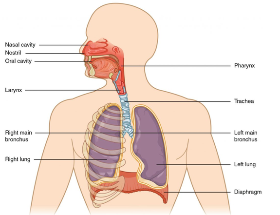
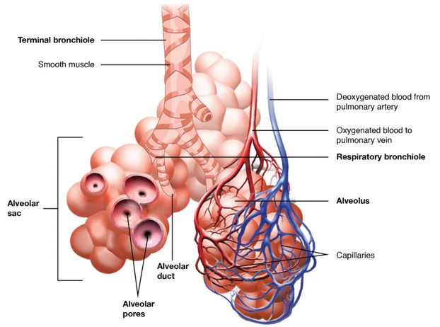
In each breath, carbon dioxide is exhaled and fresh oxygen is inhaled and passes through the air conduction zone. The structures include: respiratory muscles to move air into and out of the lungs (mainly the diaphragm), air passageways (nose, mouth, pharynx, larynx (voice box), trachea, and primary, secondary, and tertiary (etc.) bronchia, until the air reaches the respiratory zone, which includes: respiratory bronchioles, alveolar ducts, and the gas exchange surfaces of the terminal air sacs called alveoli. These are covered by capillary blood vessels.
The vascular system, under the force of the repeatedly contracting left ventricle, transports oxygen picked-up by the blood in the lungs to all the different tissues throughout the body, and then after the cells use the oxygen to produce ATP for energy, the carbon dioxide by-product from all the body tissues is returned to the right ventricle, which pumps the blood with the C02 to the lungs and is exhaled.
A variety of diseases can affect the respiratory system, such as asthma, emphysema, chronic obstruction pulmonary disease(COPD), and lung cancer. All of these conditions affect the gas exchange process in the lungs and result in labored breathing. Severe cases are terminal (deadly).
Learning Objectives
- Describe the structures of the air conduction zone
- Describe the structures of the respiratory zone
- Describe the process of air exchange across the respiratory membrane
The autonomic nervous system (ANS) controls your respiration – the breathing of air in and out of your lungs. Breathing is of vital (life or death) importance, because every cell in the body needs a continual supply of oxygen for “cellular respiration” - the process by which energy is produced in the form of Adenosine Tri-Phosphate (ATP).
For oxidative phosphorylation to occur, oxygen is used as a reactant and carbon dioxide is released as a waste product. Although oxygen is the critical gas needed by the cells, it is the accumulation of carbon dioxide in the tissues that drives your need to breathe.
In each breath, carbon dioxide is exhaled and fresh oxygen is inhaled and passes through the air conduction zone. The structures include: respiratory muscles to move air into and out of the lungs (mainly the diaphragm), air passageways (nose, mouth, pharynx, larynx (voice box), trachea, and primary, secondary, and tertiary (etc.) bronchia, until the air reaches the respiratory zone, which includes: respiratory bronchioles, alveolar ducts, and the gas exchange surfaces of the terminal air sacs called alveoli. These are covered by capillary blood vessels.
The vascular system, under the force of the repeatedly contracting left ventricle, transports oxygen picked-up by the blood in the lungs to all the different tissues throughout the body, and then after the cells use the oxygen to produce ATP for energy, the carbon dioxide by-product from all the body tissues is returned to the right ventricle, which pumps the blood with the C02 to the lungs and is exhaled.
A variety of diseases can affect the respiratory system, such as asthma, emphysema, chronic obstruction pulmonary disease(COPD), and lung cancer. All of these conditions affect the gas exchange process in the lungs and result in labored breathing. Severe cases are terminal (deadly).
An alveolar sac is a cluster of many individual alveoli that are responsible for gas exchange. An alveolus has elastic walls that stretch during air intake, which greatly increases the surface area available for gas exchange. Alveoli are connected to each other by alveolar pores, which help maintain equal air pressure throughout the alveoli and lung.
The blood-gas barrier (aka: alveolar-capillary barrier or respiratory membrane) is the key functional element of the lung – the site of O2 and CO2 exchange between the distal airspaces and the pulmonary vasculature.
The alveolar wall consists of three major cell types:
The simple (1 layer) squamous epithelium formed by type I alveolar cells is attached to a thin, elastic basement membrane. This alveolar membrane is extremely thin and borders the endothelial membrane of capillaries. Taken together, the alveoli and capillary membranes form a respiratory membrane that is approximately 0.5 mm thick.
The respiratory membrane allows gases to cross by simple diffusion, allowing oxygen to be picked up by the capillary blood for transport to the body tissues and carbon dioxide to be released into the air in the alveoli and eliminated by exhalation.
- Type I alveolar cells are the squamous epithelial (very thin, flat, surface) cells of the alveoli, which constitute 97% of the alveolar surface area. These cells are highly permeable to gas.
- Type II alveolar cells are interspersed among the type I cells and secrete pulmonary surfactant, a substance composed of phospholipids and proteins that reduces the surface tension of the alveoli, making expansion of the lung much easier – almost effortless.
- Alveolar macrophages roam around the alveolar walls. These are phagocytic (consuming) cells of the immune system that remove debris and air-borne pathogens (bacteria, “germs”) that have reached all the way into the alveoli.
The simple (1 layer) squamous epithelium formed by type I alveolar cells is attached to a thin, elastic basement membrane. This alveolar membrane is extremely thin and borders the endothelial membrane of capillaries. Taken together, the alveoli and capillary membranes form a respiratory membrane that is approximately 0.5 mm thick.
The respiratory membrane allows gases to cross by simple diffusion, allowing oxygen to be picked up by the capillary blood for transport to the body tissues and carbon dioxide to be released into the air in the alveoli and eliminated by exhalation.
Learning Module 4B: LAB
Hold your breath. Really! See how long you can hold your breath until you start to feel uncomfortable (20-30 seconds). Do not push it! You could pass-out, fall, and hit your head.
- A typical human cannot survive more than 3-4 minutes without breathing. If you try to hold your breath too long, your autonomic nervous system will take control, forcing you to inhale.

